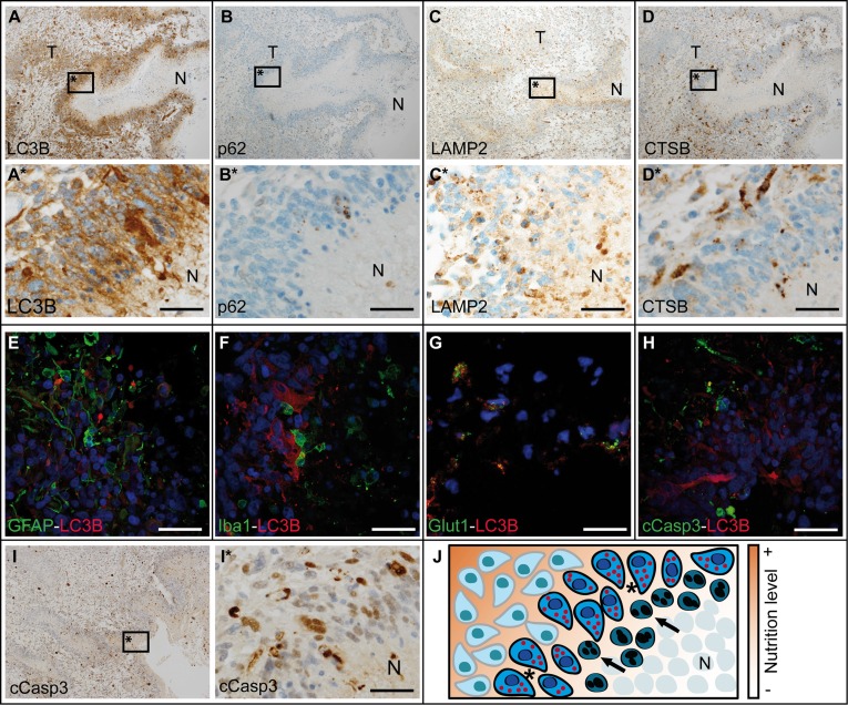
Figure 5. Autophago-lysosomal proteins are upregulated in close vicinity to necrotic foci in glioblastoma.
Overview about (A) LC3B, (B) p62, (C) LAMP2 and (D) CTSB immunohistochemistry in glioblastoma (N: necrosis, T: tumor center). (E–H) Double immunofluorescent staining against LC3B and (E) GFAP, (F) Iba1, (G) Glut1 as well as (H) cCasp3 in glioblastoma. (I) Overview of cCasp3 immunohistochemistry in glioblastoma. (A*, B*, C*, D* and I* are higher magnifications of A, B, C, D and I respectively; all scale bars: 50 μm). (J) Schematic overview of the border zone of necrotic foci with different nutrition levels in glioblastoma (arrows: apoptotic cell, *cells expressing autophagy-associated and lysosomal markers, N: necrosis).

