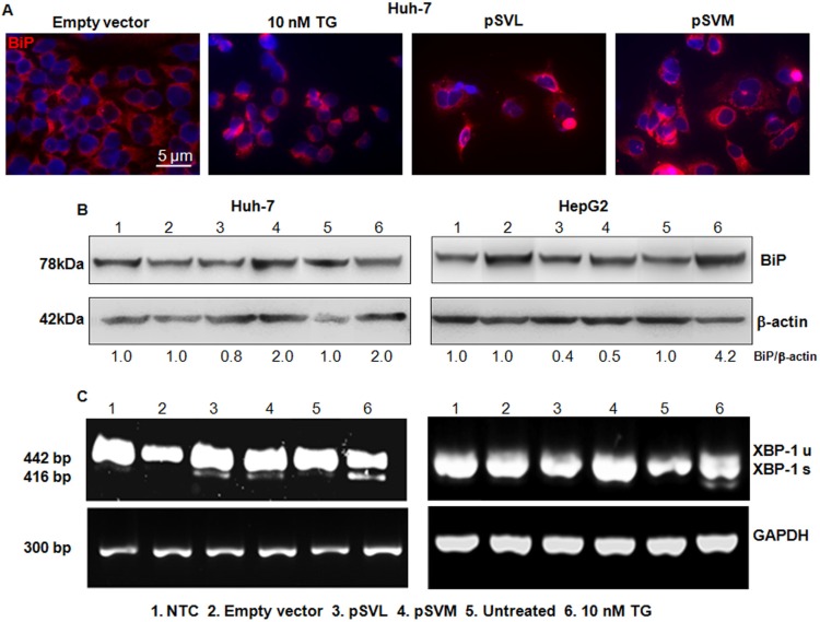
Figure 3. BIP detection and Xbp-1 splicing.
Immunofluorescence analysis of BiP after 72 hours of transfection with Empty vector, pSVL or pSVM plasmids, or treatment with 10 nM TG in Huh-7 cells (A). Immunofluorescence analysis has been performed under identical settings. Nuclei were stained with Hoechst 33342. Magnification is 630 × and scale bar represents 5 μm. Western blot results of BiP in Huh-7 (B) left blots) and HepG2 (B, right blots) cells, after 72 hours of transfection with Empty vector, pSVL or pSVM plasmids, or treatment with 10 nM TG. Densitometry results were normalized to β-actin content and are expressed relative to Empty vector for pSVL and pSVM plasmids transfected cells, and to untreated controls for TG treated cells and set in both cases at 1.0. RT-PCR analysis of total RNA isolated from Huh-7 (C) lowest left panels) and HepG2 (C, lowest right panels) cells transfected with Empty vector, pSVL or pSVM plasmids, or treated with 10 nM TG for 72 hours. For this experiment, primers amplifying both unspliced (XBP-1u, 442 bp) and spliced (XBP-1s, 416 bp) forms of XBP-1 mRNA were used. Levels of GAPDH mRNA were used as internal control.

