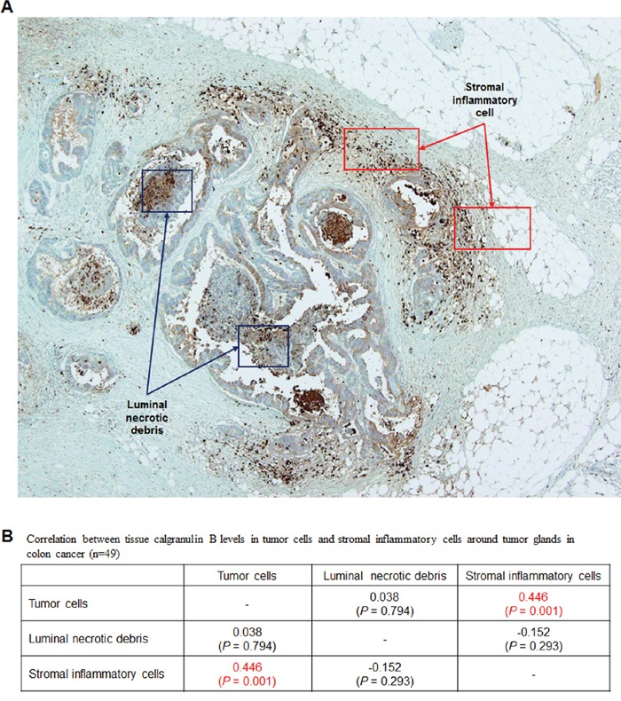
Figure 2. Evaluation of calgranulin B in colon cancer patient tumor tissues.

A. IHC analysis of calgranulin B in patient tissues. Staining was negative in all tumor tissues tested. Most positive calgranulin B staining was observed in tumor cells surrounded by inflammatory cells. B. Correlation between tissue calgranulin B levels in colon cancer tumor cells and stromal inflammatory cells around tumor glands. Calgranulin B protein level was estimated in tumor cells, luminal necrotic debris and stromal inflammatory cells (n = 49). Calgranulin B expression in colon cancer tissues was correlated with the presence of stromal inflammatory cells (Pearson correlation coefficient = 0.446, P = 0.001).
