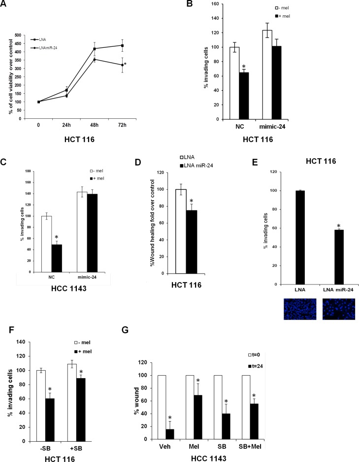
Figure 3. Melatonin impairs the capability of miR-24 to induce cell proliferation and migration.
(A) Cells were transfected with the indicated LNA and allowed to proliferate. Percentage of cells relative to control is indicated in the graph. *p < 0,05. (B) HCT 116 and (C) HCC1143 cells were transiently transfected with either NC or mimic-24 and subjected to transwell migration assays in the absence and in the presence of melatonin. Histograms show the percentage of invading cells. *p < 0,001. (D) HCT 116 cells were transfected with the indicated LNA and subjected to wound healing assay. Histograms show the percentage of wound opening. *p < 0,05. (E) HCT 116 were transfected with the indicated LNA and subjected to transwell migration assay. Histograms show the percentage of invading cells. *p < 0,01. (F) HCT 116 cells have been treated with 5 μM SB202190 for 2 hours and then subjected to transwell migration assay. *p < 0,05. (G) HCC1143 cells have been subjected to wound healing assay in the absence and in the presence of SB202190 and melatonin. Histograms show the percentage of wound opening. *p < 0,05.

