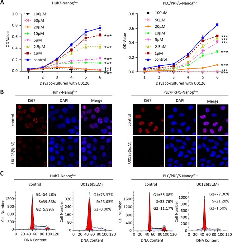
Figure 1. MEK1 inhibitor decreases liver CSCs proliferation ability in vitro.
(A) Huh7-NanogPos and PLC/PRF/5-NanogPos cells under different U0126 concentrations treatment as indicated (0 μM, 1 μM, 2.5 μM, 5 μM, 10 μM, 20 μM, 50 μM, 100 μM) were seeded (1 × 10³) and cultured for another 6 days before analyzed with CCK8. (B) Huh7-NanogPos and PLC/PRF/5-NanogPos cells were cultured with or without 5 μM U0126 for 48 hours. Cells were harvested for immunofluorescence (IF) analysis by anti-Ki67 antibodies. Scale bar, 10 μm. (C) Cell cycle profiles of 5 μM U0126 treated or DMSO treated (negative control) Huh7- and PLC/PRF/5-NanogPos cells followed by treatment with Sodium butyrate. Percentage in each histogram indicates the portion of cells remaining in each cell cycle phase.

