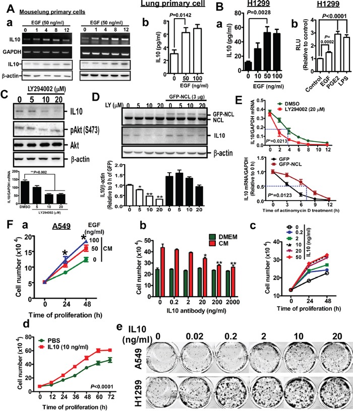
Figure 2. EGF induces IL10 expression, and IL10 increases proliferation.

A (a). After treatment with EGF for 24h in serum-free media, mouse lung primary cells were harvested for RT-PCR and Western blotting targeting IL10. (b). The medium was collected for detecting IL10 by ELISA. B (a). IL10 secretion in EGF-treated H1299 cells (b). H1299 cells-expressed pGL2-IL10 promoter were harvested for luciferase reporter assay after EGF treatment. PGE2 and LPS treatments are positive controls. Data were expressed as mean±s.e.m. C. After LY294002 treatment for 24h, H1299 cells were harvested for Western blotting to detect IL10. The result was quantified as lower panel. D. GFP-nucleolin (NCL)-overexpressed cells were treated with LY294002 for 24h, and harvested for Western blotting. Lower panel is the quantified result. Data were expressed as mean±s.e.m. (*p<0.05, **p<0.01). E. After treatment with LY294002 or GFP-NCL overexpression for 24h, cells were treated with actinomycin D for indicated time interval. Total RNA was extracted for RT-qPCR targeting IL10. F (a). A549 cells were treated with medium from cells treated with EGF for 24h, cell number was counted by hemacytometer. Data were expressed as mean±s.e.m. (*p<0.05). (b). Cells were treated with conditional media in the absence or presence of anti-IL10 antibody for 24h. Cell number was counted. (c). After treatment with recombinant IL10 for 24h, cell number was counted. (d). After 10 ng/ml IL10 treatment for indicated time, cell number was counted. (e). Cells were cultured in the presence of indicated dose of recombinant IL10 protein for 14 days. Colony was stained by methyl-blue.
