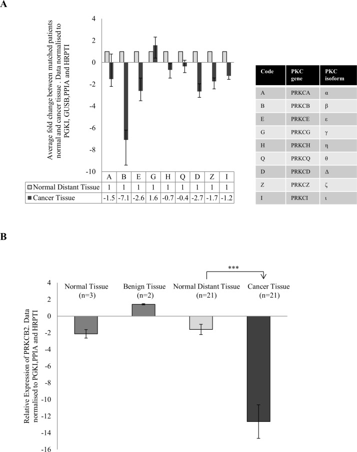
Figure 1. Gene expression of PKC genes in colon cancer.
Tissue samples measuring approximately 0.5 cm in diameter were collected from 21 patients undergoing surgery in University Hospital Limerick. Normal tissue from the 21 patients was also collected approximately 10 cm away from the cancer tissue. RNA was extracted from the tissue, cDNA was synthesised and real time PCR was carried out. All data was normalized using the housekeeping genes PRGK1, GUSB, PPIA and HRPT1. (A) Average fold change of the 9 PKC coding genes in cancer tissue of patients compared to each patients normal tissue (n = 21). Each individual patients fold change was analysed using the REST© software. The table explains the corresponding PKC coding gene and PKC isoform. (B) Gene expression of PRKCB in normal tissue (n = 3), benign tissue (n = 2) and cancer tissue (n = 21) relative to normal distant tissue (n = 21). Data was analysed using the delta delta ct method (Statistical significance based on the Welch test and Bonferroni test ***p < 0.01).

