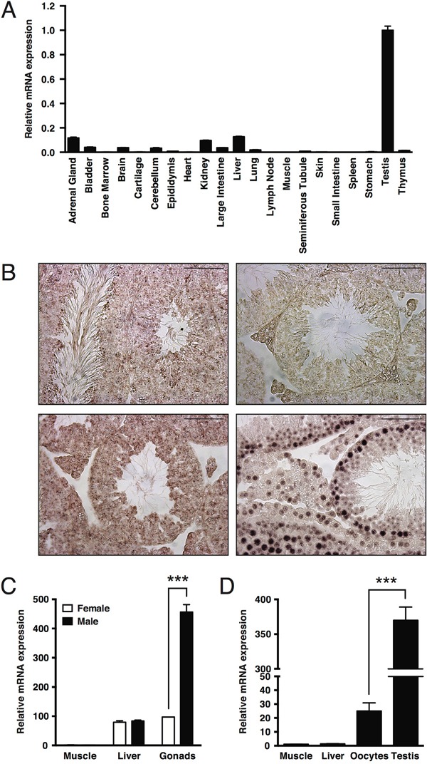
Figure 1. Elevated expression of MAPK15 in male gonads is a conserved trait in mouse and X. laevis.

A. Expression levels of Mapk15 in a panel of tissues from adult CD1 male mice, assessed by quantitative real-time PCR. B. In-situ hybridization (ISH) on paraffin-embedded adult CD1 mouse testis. Sections were probed with a Mapk15-specific LNA probe (upper left), with a LNA probe against sense miR159 from Arabidopsis thaliana (upper right) as negative control and with LNA probes against Actb (lower left) and U6 snRNA (lower right) as positive controls. Scale bars correspond to 25 μm. C. Expression levels of Mapk15 in male and female gonads from adult CD1 mice, assessed by quantitative real-time PCR. D. Expression levels of mapk15 in testis and oocytes from adult X. laevis, assessed by quantitative real-time PCR. Each bar represents the average ± SEM of three PCR replicates. Significance (p value) was assessed by Student's t test. Asterisks were attributed as follows: ***p<0.001.
