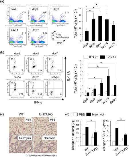
Figure 2.

Phenotypes of pulmonary γδT cells in bleomycin‐induced fibrosis. (a) Pulmonary lymphocytes were harvested from wild‐type (WT) mice (n = 3) on days 0, 3, 7, 14 and 21 after intratracheal instillation of bleomycin. Cells were stained for CD3ε, γδ T cell receptor (TCR)δ and analysed by flow cytometry. Data are representative of at least three independent experiments. Data are mean ± standard deviation (s.d.). *P < 0·05. (b) Pulmonary lymphocytes were harvested from WT mice (n = 3) on days 0, 3, 7, 14 and 21 after bleomycin exposure and stimulated by phorbol myristate acetate (PMA)/ionomycin for 6 h. Cells were stained for CD3ε, γδTCR, interferon (IFN)‐γ, interleukin (IL)−17A and analysed by flow cytometry. CD3ε+ γδTCR+ cells were gated. Data are representative of at least two independent experiments. Data are mean ± s.d. *P < 0·05. (c) Lung tissues were removed from WT (n = 3) and inrerleukin (IL)−17A–/– mice (n = 4) on day 21 after bleomycin exposure. Paraffin sections were stained with Masson's trichrome. Original magnification ×200. (d) Lung tissues and bronchoalveolar lavage fluid (BALF) were obtained from WT (n = 3) and IL‐17A–/– mice (n = 4) on day 21 after bleomycin exposure. Collagen production was determined by sircol assay. Data are representative of at least three independent experiments. Data are mean ± s.d. *P < 0·05.
