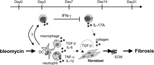
Figure 7.

Schematic diagram of the role of γδT cells in pulmonary fibrosis. Schematic diagram illustrating the role of γδT cells in the suppression of pulmonary fibrosis. After bleomycin exposure, γδT cells expanded or accumulated into lung tissues. These cells reduced pulmonary interleukin (IL)−17A+ CD4+ T cells and played regulatory roles in the pathogenesis of pulmonary fibrosis.
