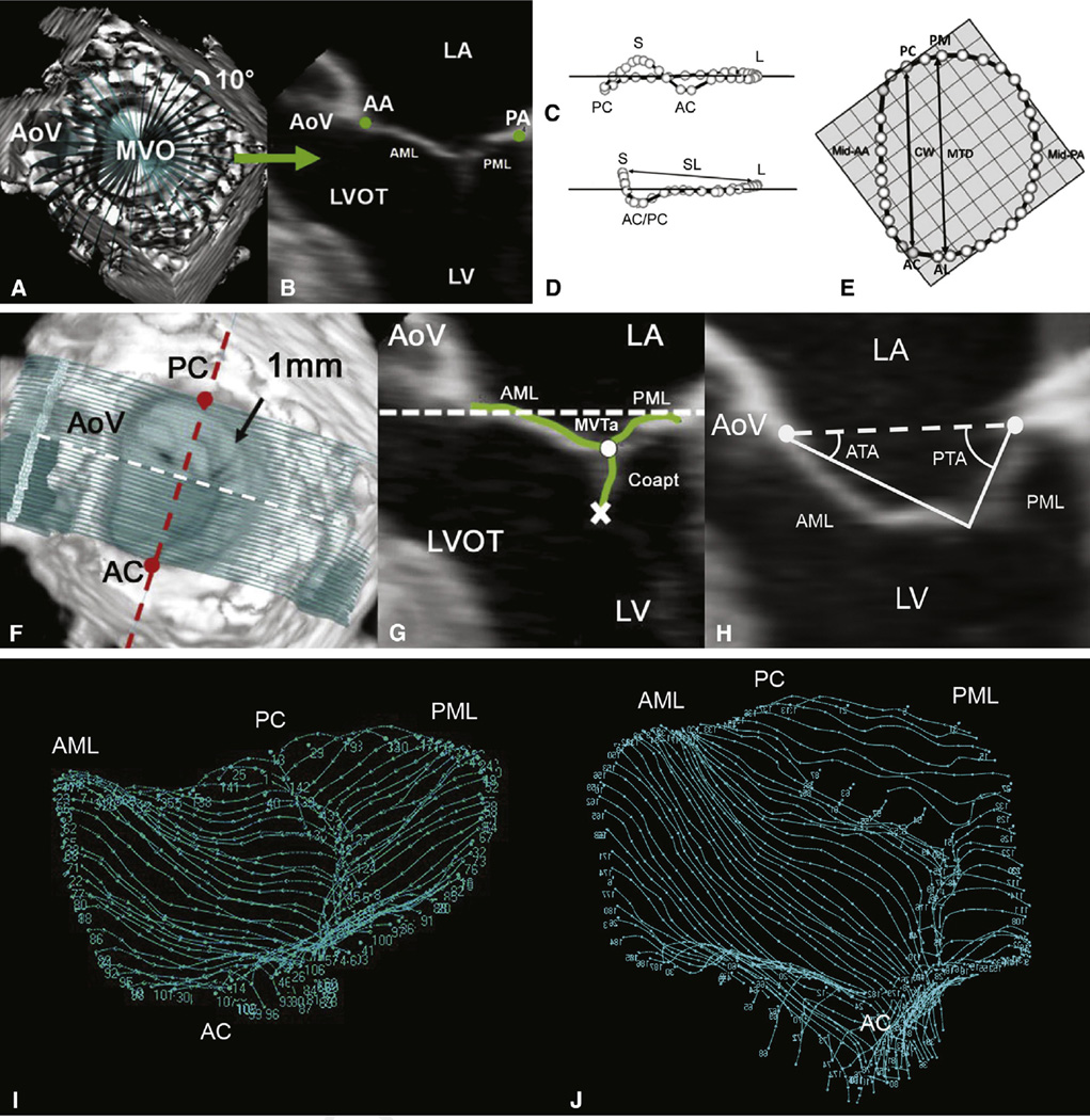
Figure 2.
3D annular (A–E) and leaflet (F–H) segmentation technique and geometric analysis. A, 3DE volume containing the mitral valve with cross-sectional planes at 10-degree increments. B, Representative 2-dimensional cross-section with green dots representing the selected annular points. Oblique (C), intercommissural (D), and transvalvular annular (E), views of a single real-time 3D–derived mitral annular model with annular landmarks and the 36 annular data points (circles). The least-squares plane has been superimposed on the annulus in each view. The least-squares plane is depicted by a horizontal line in C and D and by the check boxes in E. Determinations of septolateral diameter, intercommissural width, and mitral transverse diameter are shown in D and E. Mitral annular area (the area enclosed by the 2-dimensional projection of an annular data set onto its corresponding least-squares plane) and mitral annular circumference also were determined. F, Template of transverse cross-sections every 1 mm along intercommissural axis. G, One of the 2-dimensional cross-sections represented by the white dashed line in F; the white and red dashed lines are both within least-squares annular plane. Determinations of mitral valve tethering (MVT) area, anterior tethering angle, and posterior tethering angle are shown in G and H. The atrial surface of the mitral valve leaflets and the coaptation zone is interactively marked (green curves), resulting in a 500- to 1000-point data set for each valve. MVT area was defined as the area enclosed by the mitral annular plane (white dashed line) and the mitral leaflets for a given point along the intercommissural axis. MVT area was calculated at known intervals (0.1 mm), Δc, along the intercommissural axis. MVT volume was calculated as the sum of the incremental regional volumes (MVT area × Δcn). MVT index (MVT volume divided by mitral annular area) also was calculated for each data set. ATA and PTA were computed at known intervals (0.1 mm) along the entire length of the intercommissural axis by measuring the angle formed by the anterior or posterior leaflet tangent relative to the mitral annular plane (H). Segmental (mean) tethering angles were determined by dividing the valve into equal thirds along the intercommissural axis to conform to the standard 6 anatomic leaflet segments (A1, A2, A3; P1, P2, P3) and by calculating the mean segmental tethering angle for each specific segment on the basis of computed tethering angles at 0.1-mm intervals (along the intercommissural axis). I, Normal mitral valve. J, Tethered mitral valve. LA, Left atrium; AA, anterior mitral annulus; PA, posterior mitral annulus; AoV, aortic valve; MVO, mitral valve orifice; AML, anterior mitral leaflet; PML, posterior mitral leaflet; LVOT, left ventricular outflow tract; LV, left ventricle; S, septal aspect of the annulus; L, lateral aspect of the annulus; PC, posterior commissure; AC, anterior commissure; SL, septolateral diameter; PM, posteromedial annulus; CW, commissural width; MTD, mitral transverse diameter; MVT(a), mitral valve tethering (area); Coapt, coaptation; ATA, anterior tethering angle; PTA, posterior tethering angle.

