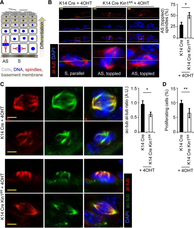
Figure 1.
Kin1 deletion in vivo results in abnormal mitosis, decreased ac-tub, and decreased cell proliferation. (A) Schematic representation of asymmetrical (AS) and symmetrical (S) cell division in the epidermis (adapted from Poulson and Lechler, 2010). Analysis of control (K14Cre) and Kin1-deleted (K14Cre Kin1fl/fl) mouse epidermis. (B) Representative images (left) of mitotic cells and quantification of asymmetrical spindles (right). (C) Representative images (left) of al-tub and ac-tub and quantification (right). (D) Percentage of phospho-histone H3-positive cells. In all graphs shown, n ≥ 3, error bars are ± SEM. P-values, t-test, *<0.05 and **<0.01.

