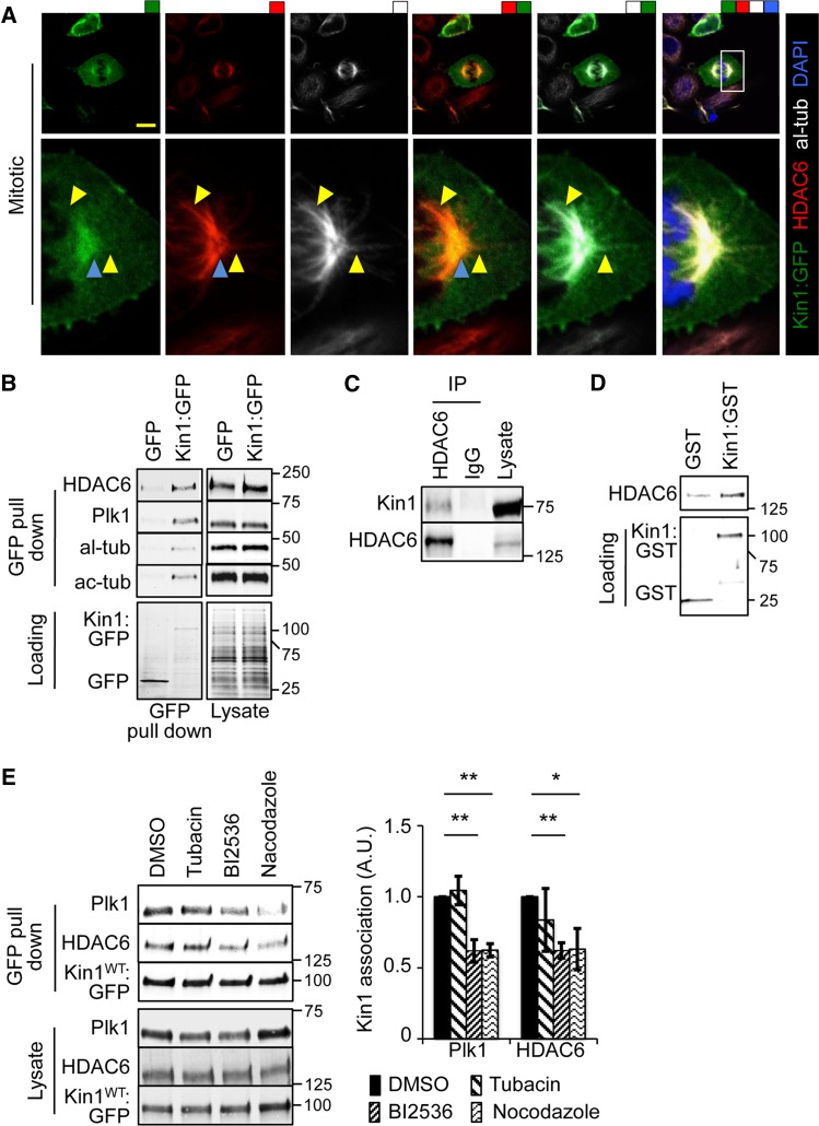
Figure 5.
Kin1 localizes with HDAC6 at centrosomes and their association is dependent on Plk1 activity and MTs. (A) Kin1:GFP co-localizes with al-tub and HDAC6 at centrosomes (blue arrowheads) and along MT (yellow arrowheads). (B) GFP pull downs from lysates of cells expressing Kin1:GFP or GFP were probed with the indicated antibodies. (C) Lysates were immunoprecipitated with anti-HDAC6 antibody and probed with Kin1 and HDAC6 antibodies. (D) In vitro binding assay of recombinant HDAC6 with either GST:Kin1 or GST. (E) GFP pull downs from lysate of cells expressing Kin1:GFP in the presence of DMSO, tubacin, BI2536, or nocodazole. In all graphs shown, n ≥ 3, error bars are ±SEM. P-values, t-test, *<0.05 and **<0.01.

