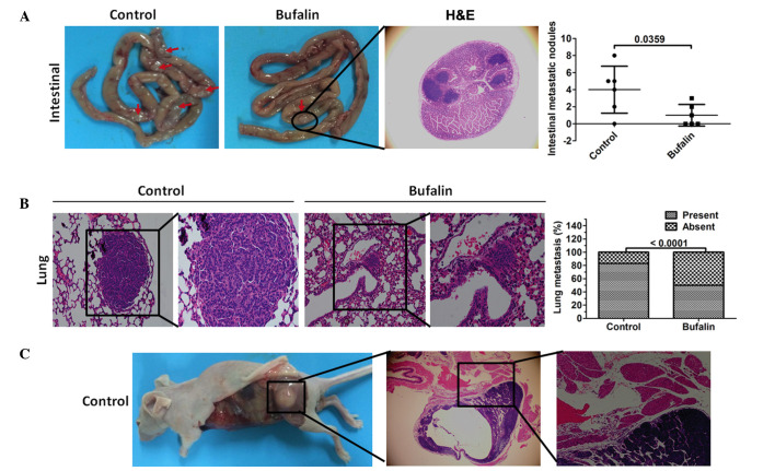
Figure 4.
Bufalin inhibits systemic metastasis of MiaPaCa2/GEM cells in vivo. A systemic metastasis model was established by injecting mice with MiaPaCa2/GEM cells pretreated with bufalin via the tail vein. (A) Representative images of intestinal metastasis. The red arrows indicate intestinal tumor lesions. Fewer intestinal lesions were observed in the bufalin-treated group. H&E staining was used to visualize the intestinal tumor lesion in the bufalin-treated mice under a microscope (magnification, ×200). The bar graph shows the number of intestinal metastases in each group. Data are expressed as the mean ± standard deviation (n=6). (B) Representative images showing H&E staining (left: magnification, ×200; right: magnification, ×400), to detect lung metastasis in the control and bufalin-treated mice. The bar graph shows the percentage of MiaPaCa2/GEM cell injections that resulted in lung metastases. (C) Representative images of muscle metastasis; H&E staining was used to confirm metastasis to the muscles (magnification, ×200). CSC, cancer stem cell; H&E, hematoxylin and eosin.

