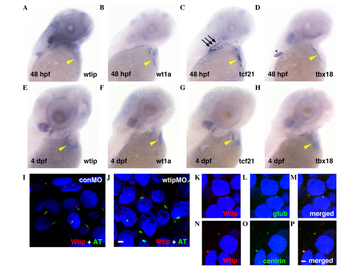
Figure 1.
wtip expression is identical to PE-specific markers during zebrafish heart development. Lateral view of (A–D) 48 hpf and (E–H) 4 dpf whole-mount in situ hybridization of (A,E) wtip, (B,F) wt1a, (C,G) tcf21 and (D,H) tbx18. At 48 hpf, (A) wtip, and PE markers (B) wt1a-, (C) tcf21- and (D) tbx18- positive cells were in the clusters near the sinus venosus, between the atrium and the yolk and near the AV junction, in contact with the ventral surface of the heart (yellow arrowhead in A–D). At 48 hpf, tcf21 expression occurred in the pharyngeal arch (black arrows in C) and tbx18 expression occurred in the pectoral fin (black asterisk in D). By 4 dpf, the wt1a-, tcf21- and tbx18-positive cells spread over the heart to cover the myocardium (yellow arrowhead in F–H). By 4 dpf, wtip-, tcf21- and tbx18-positive cells appeared to spread over the heart to cover the myocardium along the dorsal surface of the heart (yellow arrowhead in E–H). (I,J) Double immunofluorescence for the cilia marker acetylated α-tubulin (green) and Wtip (red) in confocal projections of the zebrafish PE at 48 hpf. (J) No Wtip signal was detectable in wtipMO knockdown embryos, confirming antibody specificity. (K–P, red) Localization of Wtip in the basal bodies of cilia was confirmed by double immunostaining with Wtip antibody and either (L,M, green) anti-γ-tubulin or (O,P, green) anti-centrin, which are basal body markers in heart. 4′,6-diamidino-2-phenylindole was used to counterstain nuclei (blue). Scale bar=10 µm. wtip, Wilms tumor 1 interacting protein; PE, proepicardial organ; hpf, hours post-fertilization; dpf, days post fertilization; wt1a, Wilms tumor 1a; tcf21, transcription factor 21; tbx18, Tbox 18; wtipMO, wtip morpholino oligonucleotide; conMO, control MO; AT, acetylated α-tubulin.

