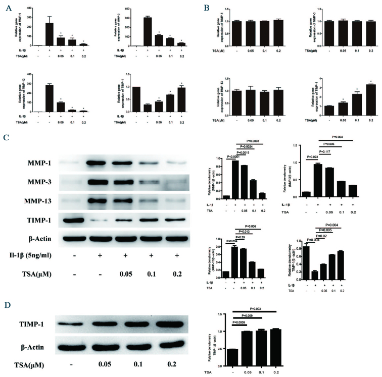
Figure 2.
Effect of TSA on MMP-1, MMP-3, MMP-13, and TIMP-1 mRNA and protein expression levels in chondrocytes. (A) Chondrocytes were pretreated with increasing concentrations of TSA for 2 h, followed by stimulation with 5 ng/ml IL-1β for 12 h. *P<0.05 vs. IL-1β alone. (B) Chondrocytes were treated with increasing concentrations of TSA for 12 h. *P<0.05 vs. untreated. Total RNA was isolated and reverse transcription-quantitative polymerase chain reaction was performed to determine relative mRNA expression levels. Levels were calculated using the 2−ΔΔCq method and expressed as the mean ± SD. (C) Chondrocytes were pretreated with increasing concentrations of TSA for 2 h, followed by stimulation with 5 ng/ml IL-1β for 24 h. (D) Chondrocytes were treated with increasing concentrations of TSA for 24 h. Proteins were harvested for western blot, and bands were quantified using densitometry and quantity one software. Data are expressed as the mean ± SD. TSA, trichostatin A; MMP, matrix metalloproteinase; TIMP-1, tissue inhibitor of metalloproteinases-1; IL-1β, interleukin-1β; SD, standard deviation.

