Figure 1.
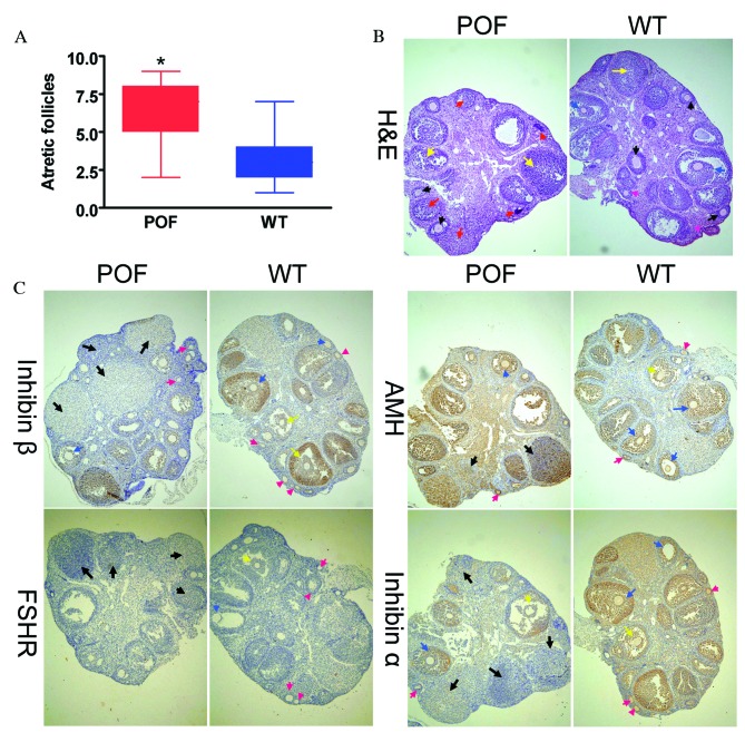
Histological analysis of ovaries. (A) The number of atretic follicles in the ovarian tissues of POF mice was significantly increased compared with control mice. *P<0.05 vs. WT; n=10. (B) H&E staining of ovarian tissues. Granulosa cells in the ovarian tissues of POF mice demonstrated clear edema and apoptosis, and increased atretic follicles. Ovarian granulosa cell injury is indicated by yellow arrows, primordial follicles by pink arrows, antral follicles by red arrows, mature oocytes by black arrows and cumulus oophorus by blue arrows. (C) Immunohistochemical staining revealed that in ovarian tissues of POF mice, the expression of the ovarian granulosa cell markers AMH, inhibinα and inhibinβ was reduced compared with those of control mice. No difference was observed in the expression of FSHR between the two groups. Black arrows indicate atretic follicles, pink arrows indicate primordial follicles, yellow arrows indicate cumulus oophorus and blue arrows indicate mature oocytes. Magnification, ×100. H&E, hematoxylin and eosin; POF, premature ovarian failure; WT, wild-type; AMH, anti-Müllerian hormone; FSHR, follicle stimulating hormone receptor.

