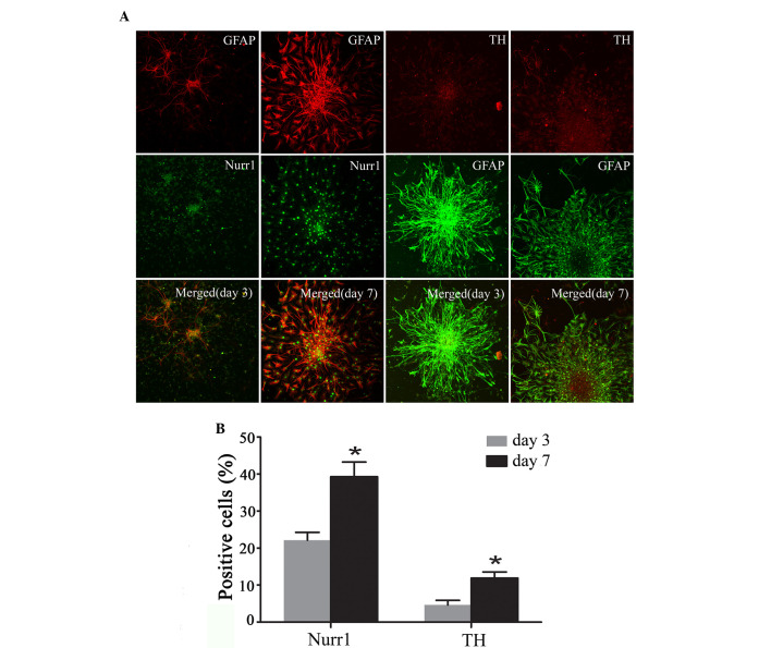
Figure 5.
Immunohistochemical analysis of expression of Nurr1 and TH and GFAP at various stages of Hip-NSC differentiation. (A) Expression of GFAP (red) and Nurr1 (green) and overlaid images at day 3 and 7; and expression of TH (red) and GFAP (green) and overlaid images at day 3 and 7 of differentiation. (B) Nurr1- and TH-positive differentiated Hip-NSCs increased significantly from day 3 (22.23±2.10 and 4.63±1.25%, respectively) to day 7 (39.30±3.96 and 11.9±1.68%, respectively). Analysis of variance was performed, followed by Dunnett's post hoc test. *P<0.05, vs. day 3. Magnification, ×200. Nurr1, nuclear receptor related-1 protein; TH, tyrosine hydroxylas GFAP, glial fibrillary acidic protein; Hip-NSC, hippocampal neural stem cell.

