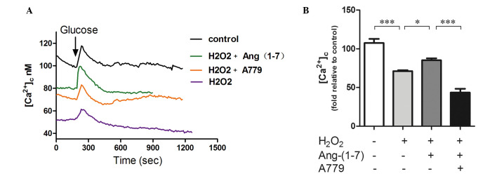
Figure 4.
Ang (1-7) restores glucose-stimulated calcium in the presence of H2O2. (A) H2O2 clearly decreased the calcium fluorescence intensity when compared with the control groups. Pre-treatment with 10−8 mol/l Ang (1-7) for 2 h prior to adding H2O2 upregulates calcium fluorescence significantly, and treatment with A779 (10−8 mol/l for 2 h) can selectively inhibit this effect. Ang (1-7) restored the amplitude of calcium (first phase of insulin secretion) in the presence of H2O2. (B) Graph of calcium fluorescence intensity. Pre-treatment with 10−8 mol/l Ang (1-7) for 2 h prior to adding H2O2 upregulates calcium fluorescence by 25% (P<0.05), and A779 (10−8 mol/l for 2 h) can selectively inhibit this effect (P<0.05). *P<0.05 and ***P<0.001. Data are presented as the mean ± standard error of the mean (n=3). Ang (1-7), angiotensin (1-7).

