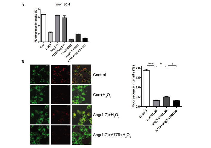
Figure 5.
Ang (1-7) restored the MMP in the presence of H2O2. (A) Flow cytometry using JC-1 in INS-1 cells. (B) JC-1 staining in INS-1 cells. MMP was evaluated by laser confocal microscopy. Ang (1-7) decreased the green fluorescence (column 1) and increased the red fluorescence (column 2) when compared with the Con + H2O2 groups. Ang (1-7) restores MMP in the presence of H2O2. The staining was quantified and presented as a graph. Ang (1-7) treatment reversed the decreases in the MMP induced by H2O2 (15 min at 250 µM H2O2), and this effect was blocked by A779. *P<0.05 and ***P<0.001. Data are presented as means ± standard error of the mean (n=3). MMP, mitochondrial membrane potential; Con, control.

