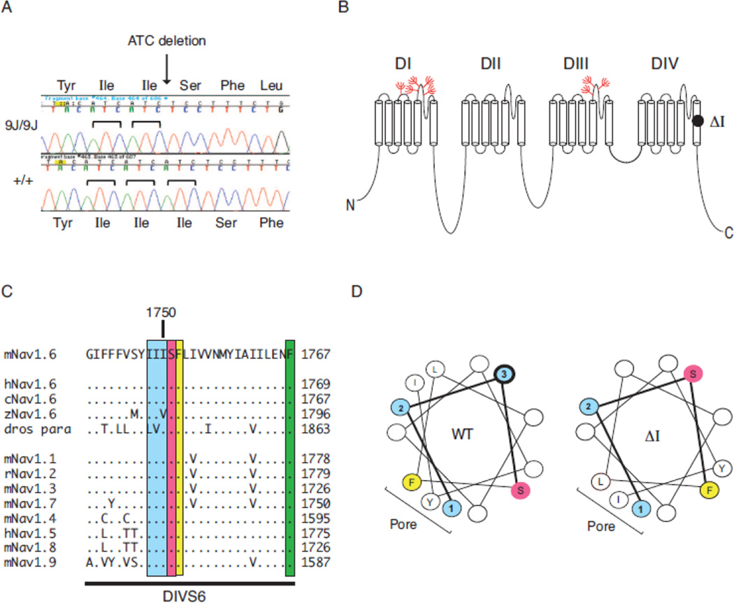
Figure 2. Deletion of isoleucine codon 1750 of Scn8a.
A) Partial sequence chromatogram of exon 27 amplified from genomic DNA of homozygous mutant and wildtype control. The 3 bp deletion in the mutant removes one of the three adjacent ATC codons. B) Location of the deleted residue in transmembrane segment DIVS6 of Nav1.6. Location of glycosylation sites (red). C) The (Ile)3 repeat is evolutionarily conserved in vertebrate sodium channels. The Drosophila homolog contains conservative substitutions of leucine and valine for two isoleucine residues. Dots represent amino acid identity. D) Helical wheel representation of transmembrane segment DIVS6 beginning with isoleucine 1748. Blue, Isoleucine residues 1748–1750 (labeled 1–3). Yellow, phenylalanine 1752; pink, hydrophilic residues serine 1751 and asparagine 1757. The image was generated with the helical wheel projection program rzlab.ucr.edu/script/wheel/wheel/cgi.

