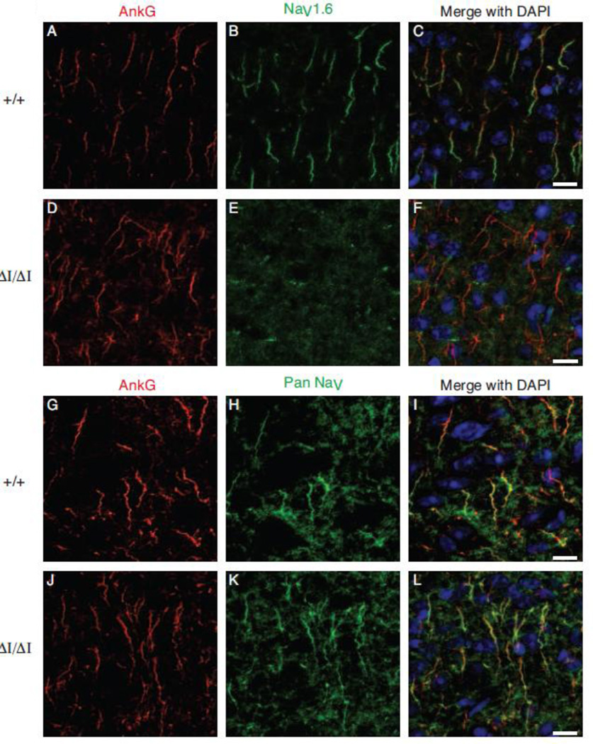
Figure 4. Nav1.6Δ1750 is not detectable at the axon initial segment (AIS).
Axon initial segments of the layer II/III of the visual cortex from 4 month old mice were immunolabeled using anti-Ankyrin G (AnkG. Red). NaV1.6 staining is localized to the distal AIS in wild-type mice (C), while AnkG-NaV1.6 co-localization was absent in ΔI/ΔI mice (F). A pan-NaV antibody (green) reveals sodium channels distributed along the length of the AIS in both WT and ΔI/ΔI. 8 micron optical sections are shown as sum intensity z-stack of 23 slices. Scale bar 10 microns.

