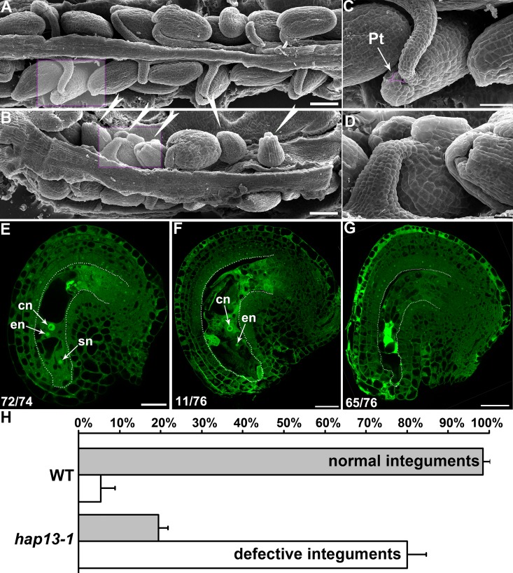
Fig 2. Ovule development in wild type and hap13-1.
(A-B) Scanning electron micrographs (SEMs) of wild-type (A) or hap13-1 (B) mature pistils at 48 hours after pollination (HAP). The defective hap13-1 ovules are highlighted with arrowheads. (C-D) Close-ups of wild-type (C) or hap13-1 (D) ovules as indicated in the shaded rectangle box in (A-B). An incoming pollen tube (Pt) at the micropyle of a wild-type ovule is false-colored pink. Two ovules with broken or incomplete outer integuments are shown in (D). (E-G) CLSM of a mature ovule from wild type (E), or from a mildly affected (F) or severely affected (G) hap13-1 mutant. Mid-optical sections of stage 3-V ovules are shown. The number of displayed type among all ovules examined is shown at the bottom left. (H) Quantitative analyses of integument development. Results in (H) are means ± standard errors (SE, N = 15). Bars = 100 μm for (A-B); 50 μm for (C); 20 μm for (D); 25 μm for (E-G).

