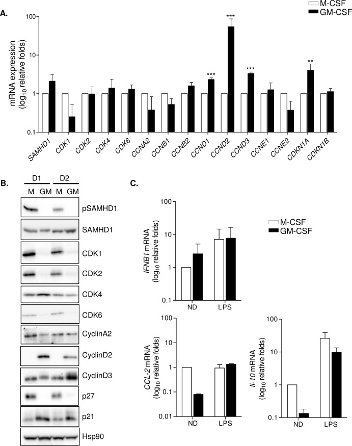
Fig 2. M-CSF and GM-CSF macrophages present differential expression of cell cycle-related proteins and SAMHD1 activation.
(A) Gene expression of cell cycle-related genes and SAMHD1 restriction pathway. mRNA levels of SAMHD1, CDK1, CDK2, CDK4 and CDK6, the corresponding cyclins (A2, B1, B2, D1, D2, D3, E1 and E2) and CDK2 inhibitor p21 (CDKN1A) was quantified in M-CSF (white bars) and GM-CSF (black bars) differentiated macrophages. Data is normalized to M-CSF relative expression. Mean ± SD of 3 independent donors is shown. ** p<0.005; *** p<0.0005. (B) Western blot showing protein expression of different cell cycle proteins, SAMHD1 expression and activation and Hsp90 as loading control. Two representative donors are shown. M; M-CSF MDM, GM; GM-CSF MDM. (C) Induction of gene expression following LPS (100 ng/ml) treatment of M-CSF and GM-CSF macrophages. Expression of IFNB1, CCL-2 and IL-10 were evaluated. Data is normalized to untreated M-CSF condition. Mean ± SD of 3 independent donors is shown.

