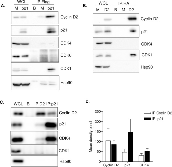
Fig 5. Co-immunoprecipitation (IP) of cyclin D2 with p21.
(A) Co-IP assay of 293T cells transfected with a plasmid expressing Flag-tagged p21. Lysates from mock- transfected (M) HEK293T cells or transfected with a Flag-p21 expression plasmid (p21) were subjected to immunoprecipitation with anti-Flag antibodies attached to sepharose or sepharose alone (B; beads). Whole cell lysates (WCL) and immunoprecipitates were analyzed by immunoblotting with an anti-cyclin D2 antibody or different CDK antibodies (CDK4, CDK6 and CDK1). Anti-p21 and anti-Hsp90 antibodies were used as controls. (B) Co-IP assay of 293T cells transfected with a plasmid expressing HA-tagged cyclin D2. As in (A) Lysates from mock- transfected (M) HEK293T cells or transfected with a HA-cyclin D2 expression plasmid (D2) were subjected to immunoprecipitation with anti-HA antibodies attached to sepharose or sepharose alone (B; beads). Whole cell lysates (WCL) and immunoprecipitates were analyzed by immunoblotting with anti-p21, anti-CDK4 and anti-CDK1 antibodies. Anti-cyclin D2 and anti-Hsp90 antibodies were used as controls. (C) Co-IP assay of endogenous cyclin D2 and p21 in GM-CSF primary macrophages. Lysates from primary macrophages where subjected to immunoprecipitation with anti-cyclin D2 or anti-p21 antibodies, attached to sepharose or sepharose attached to IgG alone (B, beads). Whole cell lysate (WCL) and immunoprecipitates were analyzed by immunoblotting with anti-cyclin D2, anti-p21, anti-CDK4 and anti-CDK1 antibodies. Anti-Hsp90 antibody was used as a control. For all Co-IP assays, a representative experiment out of 3 is shown. (D) Quantification of endogenous immunoprecipitated proteins with anti-cyclin D2 (white bars) or anti-p21 antibodies (black bars). Mean band density values ± SD of three independent donors is shown.

