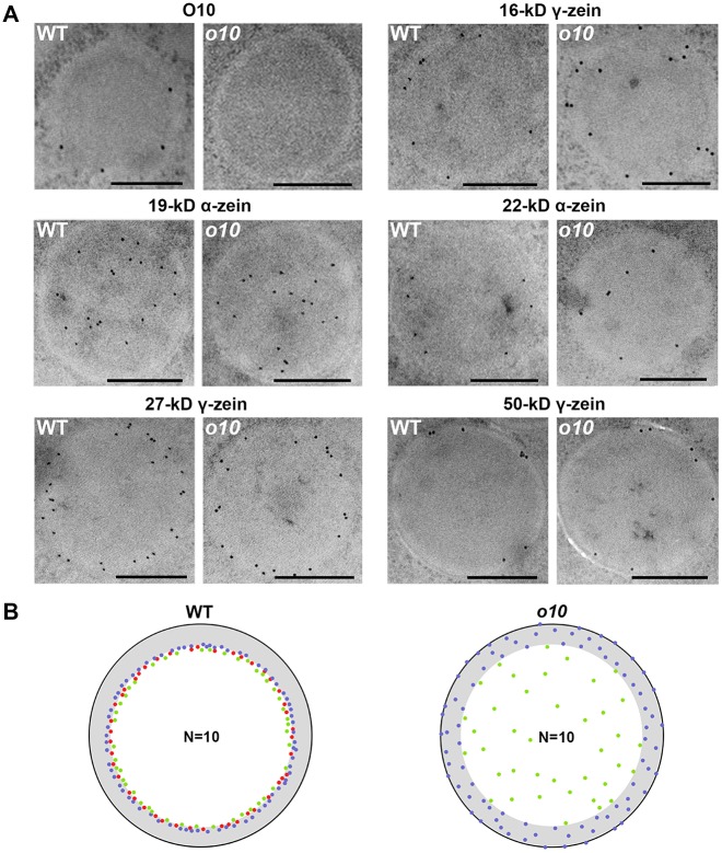
Fig 6. The distribution of different types of zeins in wild-type and o10 PBs.
(A) Immunolocalization of O10, 16-kD γ-zein, 19-kD α-zein, 22-kd α-zein, 27-kD γ-zein and 50-kD γ-zein in the PBs of wild-type and o10 at 21 DAP. Bars = 0.5 μm. (B) Ten pictures of each 16-kD γ-zein (blue dot), 22-kD α-zein (green dot) and O10 (red dot) were merged together in the same PB of wild-type and o10 according to the distance from the protein body surface to the center of the gold particle, which was measured using ImageJ.

