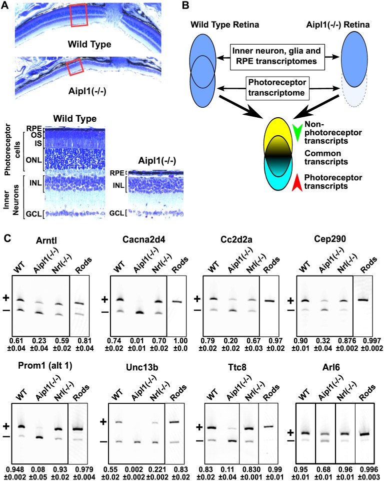
Fig 1. Identification of differentially spliced exons in photoreceptors.
(A) Retinal sections from wild type and Aipl1(-/-) mice stained with toluidine blue at postnatal day 50. Low magnification images show the overall retinal structure near the site of the optic nerve (top). Red rectangles indicate the position of the magnified images shown below. Below, high magnification images show the layered retinal structure. The Aipl1(-/-) animals lack layers formed by the photoreceptor cells: outer nuclear layer (ONL), inner segment (IS) and outer segment (OS). The retinal pigmented epithelium (RPE), the inner nuclear layer (INL) and ganglion cell layer (GCL) are intact in the Aipl1(-/-) animals. (B) Experimental approach for identifying transcripts differentially expressed in photoreceptors. The retina transcriptome is an aggregate of the transcriptomes of multiple cell types. Approximately 40–60% of the cells in the neural retina are photoreceptors. Due to the abundance of photoreceptors in the retina, their loss produces changes in the retinal transcriptome that are readily detectable. (C) RT-PCR analysis of the inclusion levels of exons identified in the RNA-Seq analysis in retina from wild type, Aipl1(-/-), Nrl(-/-) mice and flow sorted rod photoreceptors (labeled Rods). The exons include the previously described photoreceptor specific exon 2A in the Ttc8 gene and retina enriched exon 6 in the Arl6 gene. The bands corresponding to the exon skipped and exon included mRNA isoforms are labeled with ‘+’ and ‘-’, respectively. The relative exon inclusion and standard error of three independent replicates are shown below each lane.

