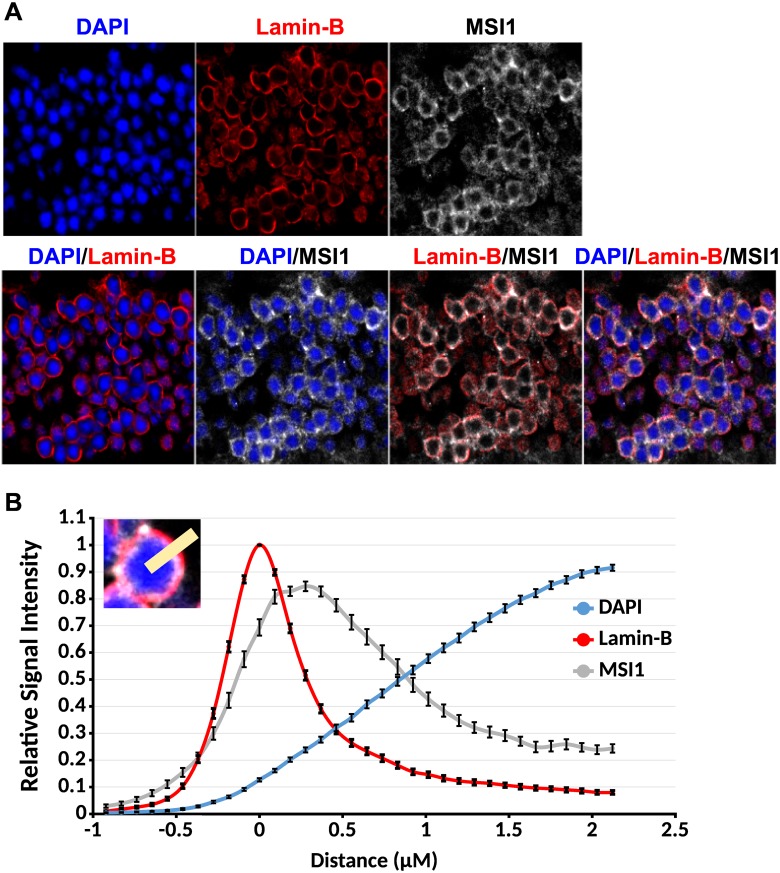
Fig 5. Musashi 1 is present in the nuclei of photoreceptor cells.
(A) Immunofluorescence staining of the outer nuclear layer on 4μm retinal sections. The nuclear envelope is stained with Lamin-B antibody (red). MSI1 staining is shown in gray. The nuclear DNA is stained with DAPI (blue). (B) Quantification of the Lamin-B, MSI1, and DAPI signal in the nuclei of photoreceptor cells. Lamin-B, MSI1, and DAPI fluorescence intensities were measured along a line perpendicular to the nuclear envelope (inset). The intensities measured on 53 nuclei were normalized and aligned to the peak of the Lamin-B staining.

