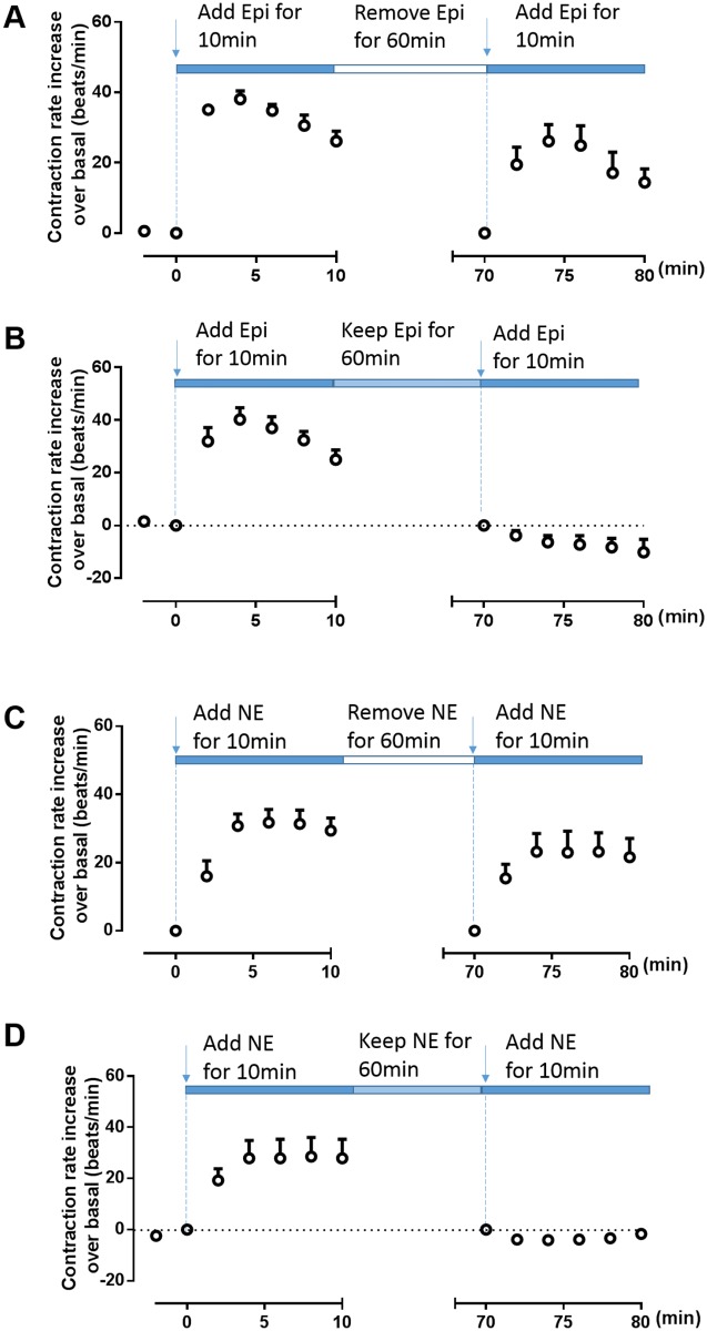
Fig 1. Cardiomyocyte contraction response to epinephrine or norepinephrine under different stimulation conditions.
Cardiac myocytes isolated from β1AR knockout mice that express endogenous β2AR were stimulated with 10 μM epinephrine (Epi) or norepinephrine (NE) for 10 min and then re-challenged with (B, D) or without (A, C) Epi or NE for 60 min. The Epi- (A, B) or NE- (F, H) induced contraction rates were recorded. Data are shown as means ± SDs from three independent experiments.

