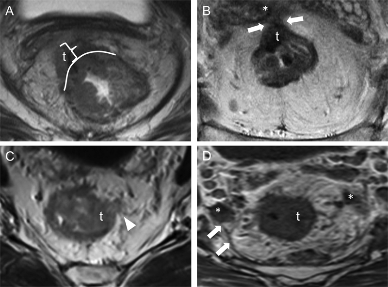
Fig. 2.

High-risk rectal cancer features on MRI. Transaxial T2WIs from four different patients show various high-risk primary tumor features. In (A), a right anterolateral rectal tumor (t) extends beyond the external aspect of the rectal wall (white curved line) into the mesorectal fat, with an extramural depth of greater than 5 mm (bracket). In (B), an anterior rectal tumor (t) extends through the mesorectal fat to involve the mesorectal fascia (arrows) and the right seminal vesicle (asterisk); these findings are compatible with T4 disease. In (C), a left lateral rectal tumor (t) exhibits extramural vascular invasion (arrowhead); the serpiginous hypointense structure represents tumor within the vessel lumen. In (D), a left lateral rectal tumor (t) with spiculations extending into the perirectal fat is associated with several enlarged lymph nodes (asterisks) that abut the mesorectal fascia (arrows), resulting in a circumferential resection margin of < 1 mm. MRI, magnetic resonance imaging; T2WI, T2-weighted images.
