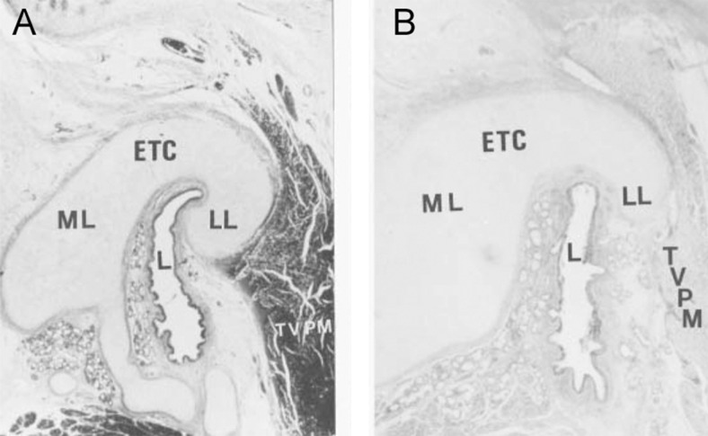
Fig. 3.
Photomicrograph of cross sections through the midcartilaginous portion of the Eustachian tube: a control case (6-week old female) and b cleft palate case (7-week old male). The photomicrographs show differences in curvature of the lumen and cross-sectional area of the Eustachian tube development of the cartilage between the normal child and the child with a cleft palate (hematoxylin-eosin stain). ETC Eustachian tube cartilage, L Eustachian tube lumen, LL lateral lamina of Eustachian tube cartilage, ML medial lamina, TVPM tensor veli palatini muscle. (Reproduced with permission from Matsune. [19])

