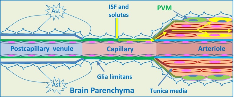
Fig. 3.
Intramural perivascular drainage of interstitial fluid (ISF) out of the brain parenchyma. As an arteriole loses its tunica media, it becomes a capillary and then a post-capillary venule. Astrocyte end feet closely invests the surface of capillaries and form the glia limitans. Capillary walls are the site of the blood–brain barrier (BBB) for solutes; the glial and endothelial components of the basement membrane (green) are fused. The wall of the post-capillary venule is the BBB for lymphocytes and other inflammatory cells to cross from blood to CNS (see Fig. 6); the glial (blue) and endothelial (green) basement membranes are not fused. Interstitial fluid (ISF) and solutes drain from the extracellular spaces in the brain parenchyma through gaps between astrocyte end feet (yellow arrow) to enter bulk flow channels in basement membranes of cerebral capillaries. From there, ISF drains into basement membranes between smooth muscle cells in the tunica media of arterioles and arteries (yellow arrows): this is the intramural perivascular drainage pathway. Tracers following this pathway are taken up by smooth muscle cells in the tunica media and by perivascular macrophages (PVM) on the outer aspects of arterioles and arteries. Neither the endothelial basement membrane of arterioles and arteries (light blue) nor the outer basement membranes of the artery wall (green) are involved in the intramural perivascular drainage of ISF from the CNS

