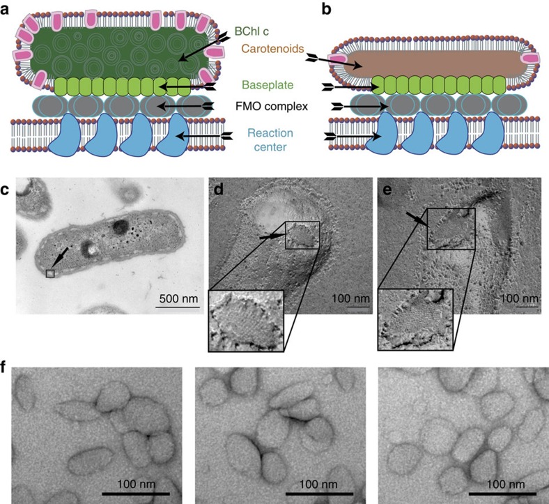
Figure 1. Organization and structure overview of the chlorosome and the carotenosome.
(a) Cartoon of a chlorosome. (b) Cartoon of a carotenosome showing CsmA (green), non-CsmA polypeptides (magenta), the surrounding lipid monolayer (red), BChl a rods (dark green), carotenoids (brown), the FMO complex (grey) and the reaction centres (dark blue) in the cytoplasmic membrane. (c) Plastic (epon) embedded thin section of the whole bacterium Cba. tepidum, arrow points to a boxed chlorosome. Baseplates of freeze fractured Cba. tepidum cells on a (d) wild-type chlorosome, (e) bchK mutant, that is, a carotenosome (zoom shows higher resolution part of the images). (f) Isolated carotenosomes prepared by negative stain embedding with uranyl acetate (Supplementary Note 1 and Supplementary Figs 1 and 2).

