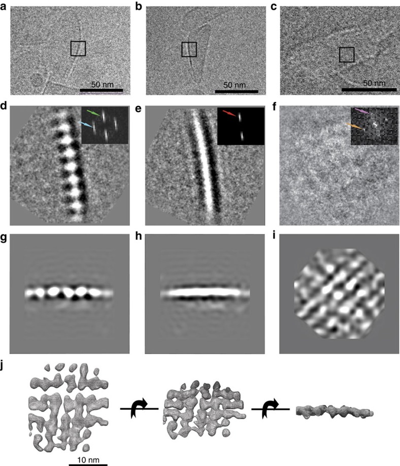
Figure 2. Carotenosome structure organization by cryo-EM.
(a–c) Cryo-EM examples of ice-embedded carotenosomes that contains characteristic features of the baseplate. (d–f) 2D class-averages from the three different views taken to represent two side views and one top view, all 90° apart: (d) string of ‘beads' (e) single stripe view, (f) weak-contrast mesh-like view (box size is 30.7 nm). Insets: Power spectra indicating repeat directions and distances in the class-averages. Green arrow corresponds to 47 Å and represents the thickness of the baseplate, whereas the blue arrow corresponds to 33 Å, indicating a specific internal repeating distance in one direction and red arrow revealing a repeating distance of 41 Å in another direction. Orange and purple arrows reveal repeating distances of 33.8 and 35.7 Å respectively. (g–i) Three perpendicular projections of the refined 3D model. (j) Electron density model showing the density map as a mesh wireframe (Supplementary Fig. 8b).

