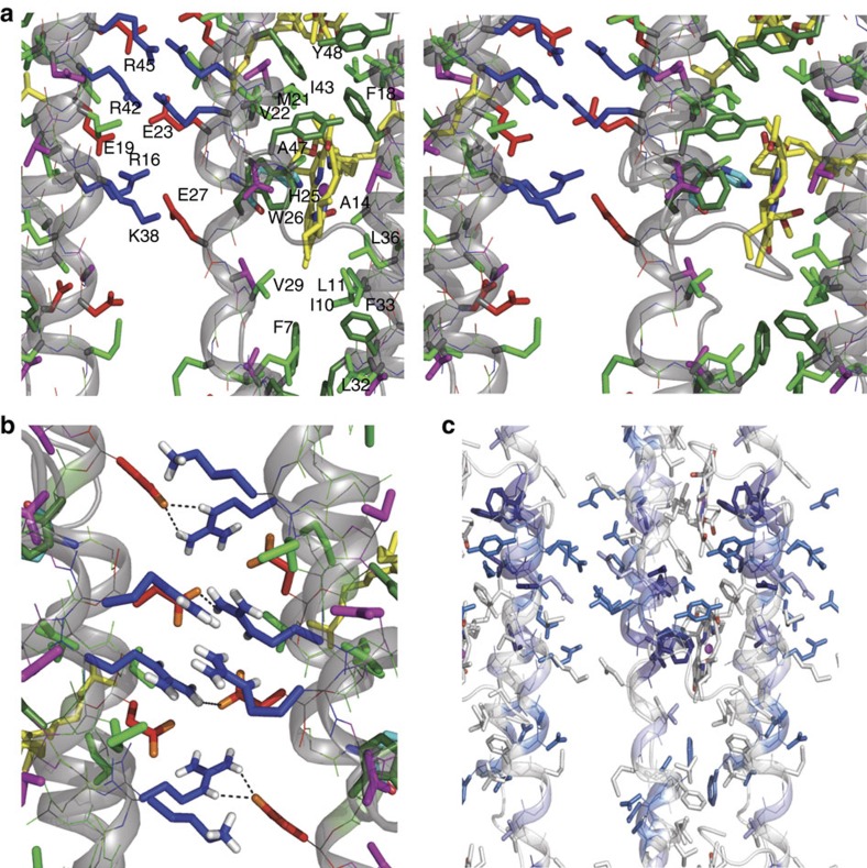
Figure 5. Details of the structure of the CsmA baseplate.
(a) Stereo view of details in the baseplate structure, see legend to Fig. 4. (b) Side chain hydrogen bonding in CsmA baseplate structure. H atoms are shown for polar side groups, O atoms are shown in orange. Hydrogen bonds are highlighted with black dashed lines. (c) Molecular structure of the CsmA baseplate visualizing homologous residues; fully conserved residues: dark blue, strongly similar residues: light blue, weekly similar: pale blue. The aligned species and corresponding sequences are shown in Supplementary Table 2.

