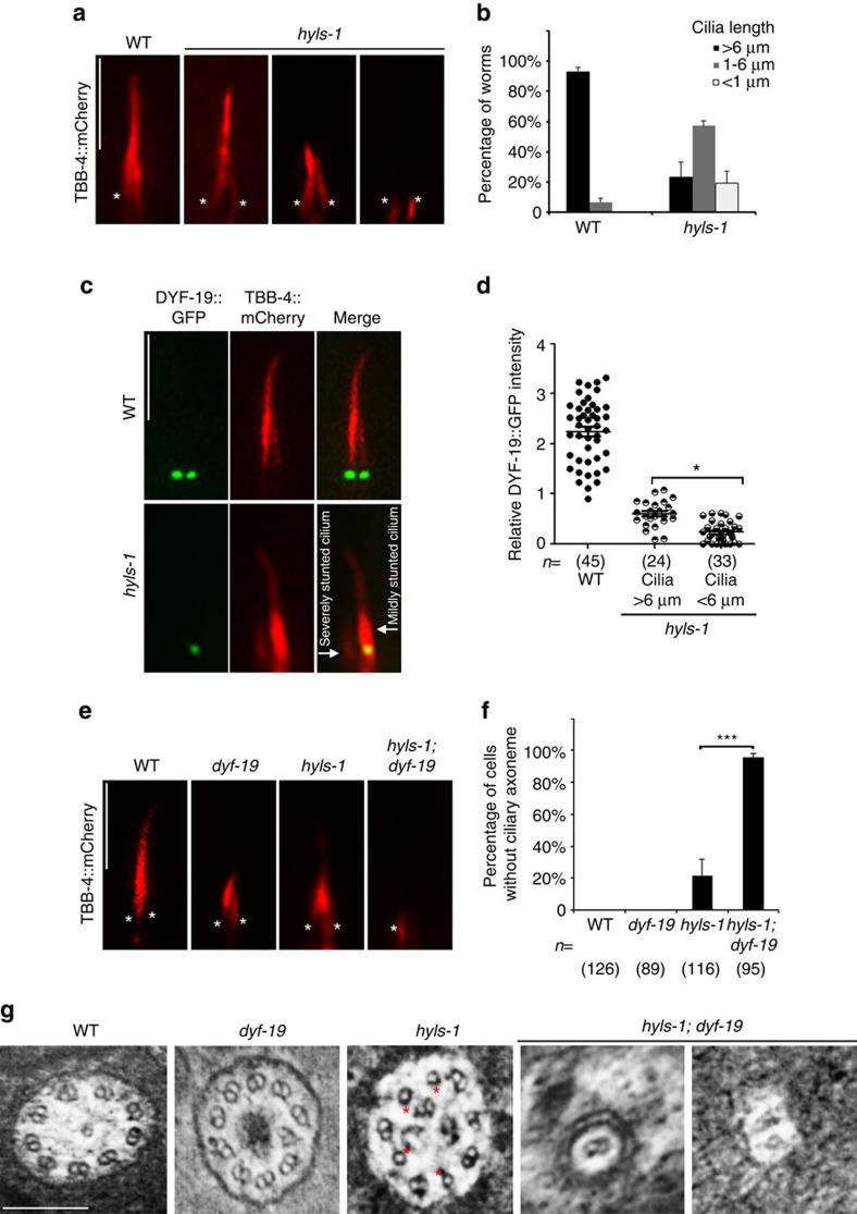
Figure 3. Additional deletion of DYF-19 in hyls-1 mutants completely abrogates ciliogenesis.
(a) hyls-1 mutants exhibit variable defects in axoneme elongation. Phasmid cilia were labelled with the mCherry-tagged axoneme marker β-tubulin TBB-4. Asterisks indicate cilia base. (b) Quantification of cilia length in WT and hyls-1 mutants. Average of three independent experiments. n>100 cilia for each genetic background in each experiment. (c) Representative images of phasmid cilia co-labelled with mCherry-tagged TBB-4 and GFP-tagged DYF-19. The severity of cilia truncation in hyls-1 mutants is correlated with the level of residual DYF-19 at the cilia base. (d) Quantification of DYF-19 signal in WT and hyls-1 mutants. n represents the number of cilia analysed. (e) Additional deletion of DYF-19 in hyls-1 mutants completely abrogates ciliogenesis. Cilia are labelled with TBB-4. Asterisks indicate cilia base. (f) Quantification of phasmid neurons without visible axoneme based on OSM-6::GFP in WT, hyls-1, dyf-19 and hyls-1; dyf-19 double mutants, respectively. n represents the number of phasmid neurons analysed. (g) TEM analysis of the proximal axoneme (immediately distal to the TZ) of amphid neurons. Compared with WT and dyf-19 axonemes that consist of nine doublet microtubules, hyls-1 mutants frequently display missing B-tubules (red stars). dyf-19; hyls-1 double mutants show severely compromised and stunted axonemes that possess only one or two microtubules and immediately terminate only one TEM section (∼80 nm) beyond the TZ. Scale bars, 200 nm (g), 5 μm (a,c,e). Error bars indicate s.d. Student's t-test indicates significant differences; *P<0.01 and ***P<0.001.

