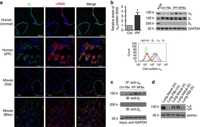
Figure 3. Lung myofibroblasts demonstrate increased α6-expression.
(a) Frozen lung tissue sections obtained from failed normal human donors, patients with IPF, saline-treated mice and bleomycin-treated mice were double-stained for α6 (green) and αSMA (red). Nuclei were stained by DAPI (blue). Confocal immunofluorescent images were overlaid to show α6-expression in αSMA-positive lung myofibroblasts. Scale bar, 50 μm; scale bar, 20 μm for mouse with bleo images. (b) Comparison for α6-expression in lung (myo)fibroblasts isolated from patients with IPF (n=10) and non-ILD control human subjects (n=6) by immunoblot; Relative levels of α6-protein normalized to GAPDH expression. Results are the means±s.d. Representative blots for α6-expression as well as β1- and β4-expression were shown. A549 cells were used as positive control for β4-expression in immunoblot analysis. Relative levels of α6-, β1- and β4-expression on the cell surface of IPF lung myofibroblasts were analysed by flow cytometry. (c) Detection of α6β1- and α6β4-complexes in IPF lung myofibroblasts by immunoprecipitation and immunoblot. (d) Identification of α6A and α6B expression in human and mouse lung tissues and fibroblasts by immunoblot; *P<0.05, one-way analysis of variance.

