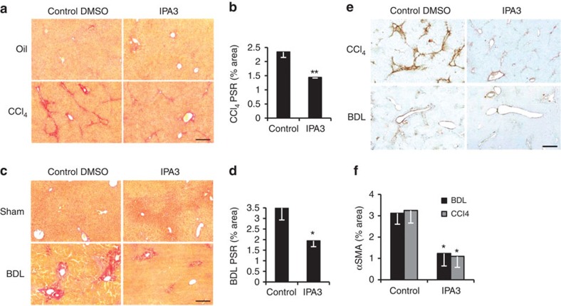
Figure 6. Pharmacological inhibition of PAK-1 improves liver fibrosis in rodents.
(a) PSR staining (collagen deposition in red) in olive oil control (Oil; top) or chronic CCl4-induced fibrosis (bottom) with DMSO (n=5) or IPA3 (n=4) treatment. (b) Quantification of surface area covered by the PSR staining in a. (c) PSR staining (collagen deposition in red) in sham-operated mice (Sham; top) or BDL to induce peribiliary fibrosis (bottom) with control DMSO (n=6) or IPA3 (n=7) treatment. (d) Quantification of surface area covered by the PSR staining in c. (e) Immunohistochemistry for α-SMA (brown; activated HSC/myofibroblast marker) in mouse livers following CCl4 (top) or BDL (bottom) induced fibrosis. (f) Quantification of surface area covered by α-SMA staining was reduced following IPA3 treatment in both models (n=5 for all animal groups). Scale bar, 500 μm. Two-tailed unpaired t-test was used for statistical analysis. Data are shown as means±s.e.m. *P<0.05, **P≤0.01.

