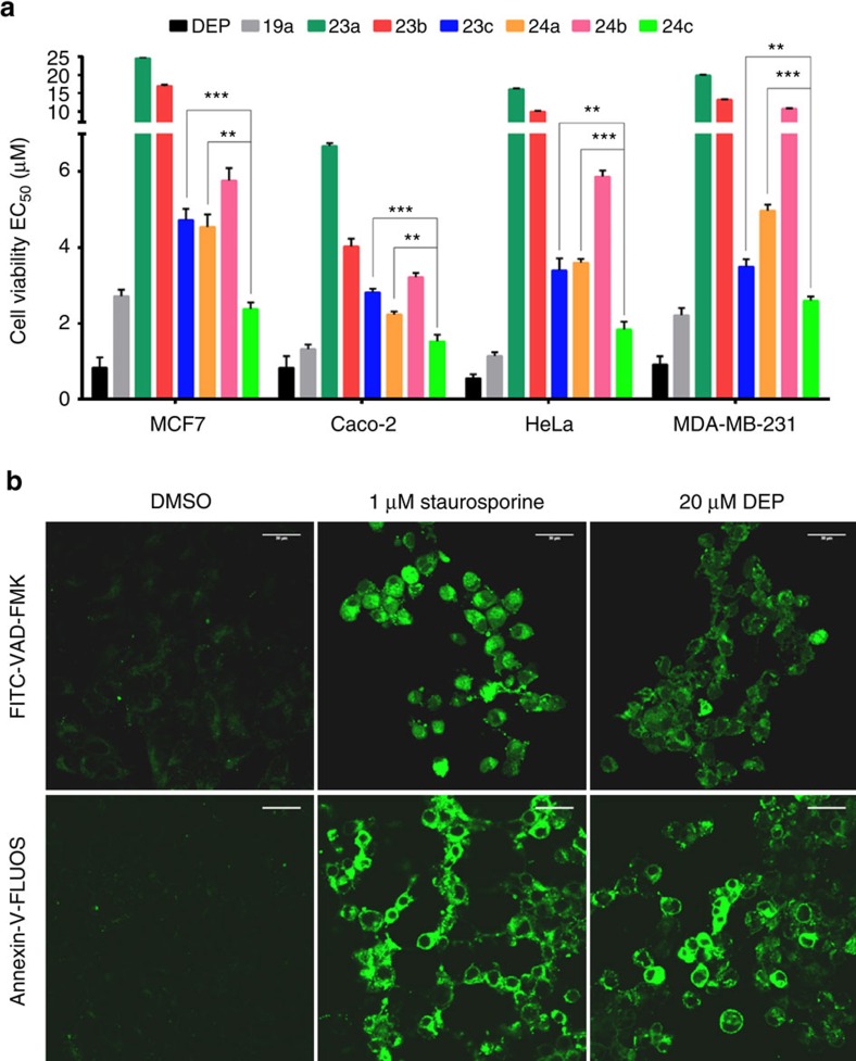
Figure 5. Assessing the cytotoxicity of deoxyelephantopin (DEP) and analogues.
(a) Cell viability measurements in four different cancer cell lines. Cells were treated with compounds for 3 days, the nuclei were stained with DAPI and counted (EC50 values±s.d. shown; n=3). **P<0.005 and ***P<0.0005 by two-sided Student's t-test. (b) Detection of caspase activation and apoptosis in MCF7 cells using FITC-VAD-FMK fluorescent probe (top) or Annexin V staining (bottom). The cells were treated with 1 μM staurosporine (positive control) or 20 μM deoxyelephantopin for 8 h. Confocal microscopy images are shown. EC50, effector concentration for half-maximum response. Scale bars, 30μm.

