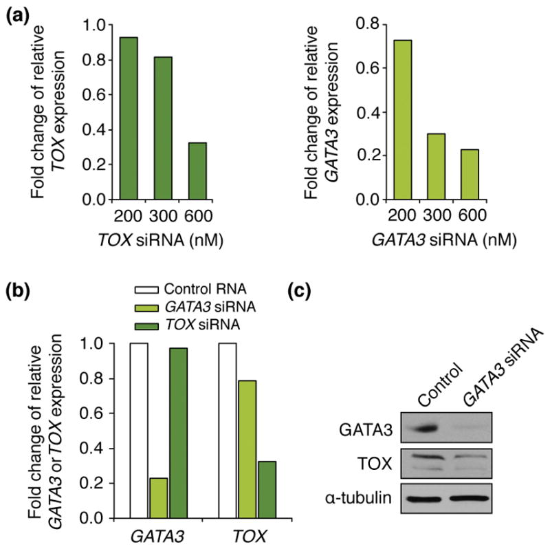
Figure 2.

GATA3 reduced TOX expression. A) Hut-78 cells were transfected with the indicated concentrations of TOX or GATA3 siRNA, and 24 hours post-transfection TOX or GATA3 mRNA levels were evaluated by qRT-PCR. Values are relative to β-actin levels. B) Hut-78 cells were transfected with 600nM of TOX or GATA3 siRNA or control RNA and were harvested 24 hours later. TOX or GATA3 mRNA levels were evaluated by qRT-PCR and are relative to β-actin levels. C) Hut-78 cells transfected with GATA3 siRNA or control RNA (CTRL) were harvested 24 hours post-transfection. Western blots of whole cell protein lysates were performed for the proteins indicated.
