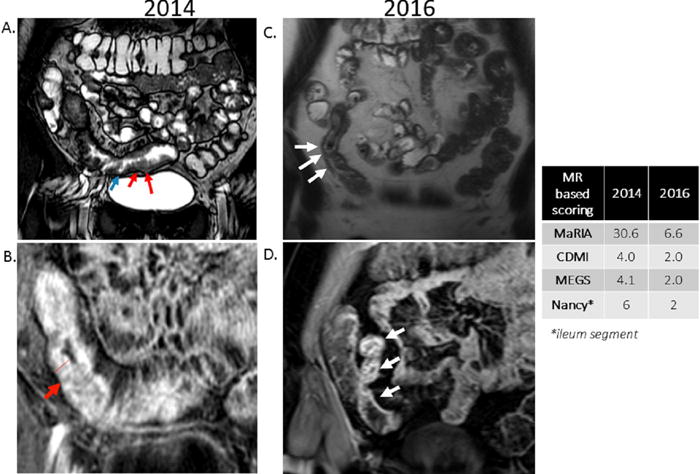
Figure 1.

29 y/o female with CD, with MRE performed in 2014 and then subsequently in 2016 (after treatment with adalimumab 40 mg subcutaneously every 2 weeks).
A. 2014 Coronal Fast Imaging Employing Steady-state Acquisition sequence demonstrating distal ileum including terminal ileum with moderate wall thickening (red arrows) and ulcerations (blue arrows).
B. 2014 Coronal post-contrast LAVA sequence demonstrating distal ileum including terminal ileum with moderate wall thickening (10.4mm), and mucosal hyperenhancement (red arrow) consistent with active Crohn’s disease.
C. 2016 Coronal T2 Half Fourier Acquisition Single Shot Turbo Spin Echo sequence demonstrating thickening (4.4mm) of a segment of ileum (white arrows), less than previous MRE in 2014.
D. 2016 Coronal T1 volumetric interpolated breath-hold fat saturated post gadolinium sequence demonstrating bowel wall hyperenhancement (white arrows) much less pronounced than previous MRE in 2014.
