Figure 4.

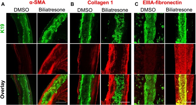
Biliatresone causes monolayer disruption, patchy obstruction, myofibroblast activation, and fibrosis in neonatal EHBD explant cultures. Neonatal mouse EHBDs were incubated for 24 hours with biliatresone or vehicle (DMSO) and immuno stained for the cholangiocyte marker K19 (green) and (A) the myofibroblast marker α‐SMA (red) and the fibrosis markers (B) collagen 1 (red) or (C) EIIIA‐fibronectin (red). K19 staining demonstrates ductal disruption, whereas the other stains demonstrate myofibroblast accumulation and matrix deposition in the submucosal and other periductal regions with biliatresone treatment. Images are representative of (A) seven independent experiments with 13 ducts for each condition, (B) three independent experiment with six ducts for each condition, and (C) three independent experiments with six ducts for each condition. Scale bars: 100 μm.
