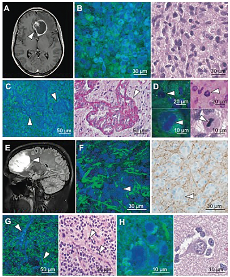
Fig. 2.
SRS and traditional microscopy of intrinsic brain tumors. A: SRS imaging of a glioblastoma multiforme (arrowhead) demonstrating ring enhancement on MRI. B: Hypercellularity and nuclear atypia of viable tumor is apparent on both SRS (left) and H & E (right) microscopy. C: Microvascular proliferation creates tortuous vascular complexes evident on SRS microscopy (left, arrowheads) and is highlighted with periodic acid-Schiff staining (right, arrowhead). D: Mitotic figures are also visible (arrowheads) with SRS microscopy (left) and H & E staining (right). E and F: A nonenhancing, low-grade oligodendroglioma (arrowhead, E) consists of hypercellular tissue with nests of “fried-egg” morphology (arrowheads, F) causing minimal axonal disruption on SRS imaging (left), as confirmed through neurofilament immunostaining (right). G and H: “Chicken wire” blood vessels (arrowheads, G) imaged with SRS (left) and H & E (right) microscopy, and perineuronal satellitosis is visible in both SRS (left) and H & E (right) microscopy (H). From Ji M, Lewis S, Camelo-Piragua S, Ramkissoon SH, Snuderl M, Venneti S, et al: Detection of human brain tumor infiltration with quantitative stimulated Raman scattering microscopy. Sci Transl Med 7:309ra163, 2015. Reprinted with permission from AAAS.

