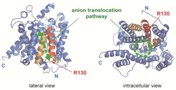
Fig. 1.
A prestin model. A structural model for the transmembrane region of prestin generated by Phyre2 is shown. The N- and C-terminal cytosolic domains are not shown in this model. The SulP and Saier motifs [13, 48] are highlighted in red and orange, respectively. Red arrows indicate the location of R130. A previously proposed anion translocation pathway [13] is also shown in green. “N” and “C” indicate the truncated N- and C-termini of the model.

