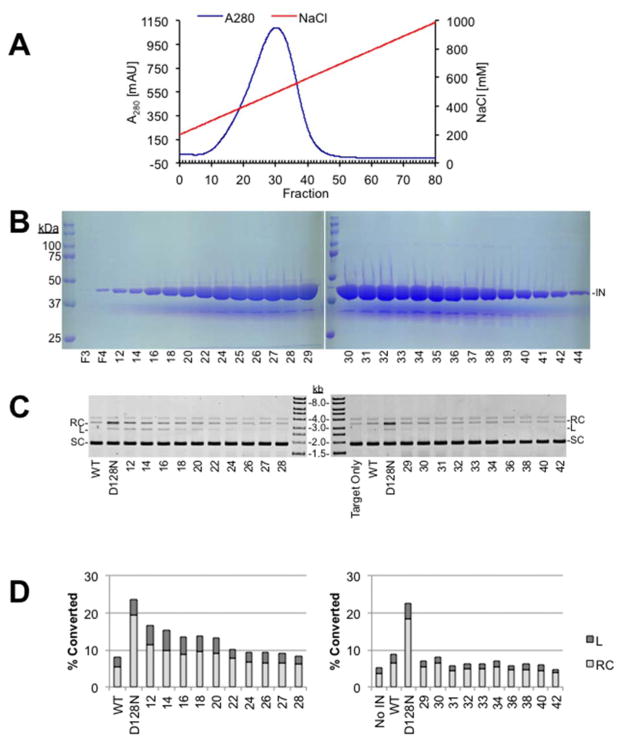
Fig. 2. Heparin sepharose chromatography of PFV IN(D128N).
A. Chromatography profile of PFV IN(D128N) fractionation with heparin sepharose. Blue line indicates the A280. Red line indicates the gradient of NaCl. B. Coomassie stained PAGE analysis of selected fractions following heparin sepharose chromatography of PFV IN(D128N), 44 kDa (IN). F3 is the void volume and F4 is the wash volume. Fraction numbers are indicated at the bottom of the gel images. C. Nuclease assay of heparin sepharose fractions separated by agarose, stained with ethidium bromide, and imaged with a Typhoon laser scanner. Relaxed circles (RC), linear (L), and supercoiled (SC) plasmid are indicated. Nuclease free PFV IN wild type (WT) is included as a negative control for nuclease activity. A previous purification of PFV IN(D128N) that did not exclude the co-purifying nuclease is included as a positive control for nuclease activity (D128N). D. Quantitation of the nuclease assay. Relative relaxation or linearization of the total plasmid associated with each heparin sepharose fraction is shown.

