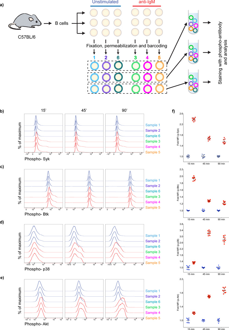
Figure 3.
Ab-based barcoding is effective in standardizing experiments using phospho-flow. (a) Purified mouse splenic B cells were divided into three sets of six samples each for each of three time points that were either unstimulated or stimulated with 10 µg/ml anti-IgM. Fifteen, 45 and 90 min later samples were fixed, permeabilized barcoded and pooled as shown. Pooled samples were stained with specific Abs and analyzed in flow cytometry. (b–e) Time-dependent changes in phosphorylation of Syk (b), Btk (c), p38 (d) and Akt (e) are shown as histogram overlays for both unstimulated cells (blue histograms corresponding to samples 1,2,6) and anti-IgM stimulated cells (red histograms corresponding to samples 3,4,5). f) Fold MFI with time after anti-IgM stimulation (red squares) and unstimulated controls (blue triangles). Each symbol represent one sample of the barcoded replicates. Error bars indicate the standard deviation. Results are representative of more than three independent experiments.

