Figure 1. Forms of AMD, (see appendix).
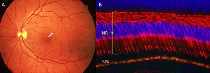
Figure 1.1: Normal Human Retina
A) Posterior pole view of a normal human retina. The fovea is arrowed. B) Immunofluorescent image of macular cones (labelled red), cell nuclei are blue (DAPI) and RPE (orange) due to autofluorescence. NR: neuroretina, RPE: retinal pigment epithelium (Courtesy of Dr Ann Milam, Scheie Eye Institute, University of Pennsylvania Philadelphia, USA)
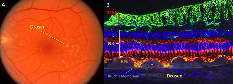
Figure 1.2: Drusen in AMD
A) Fundus view of a retina with drusen (white deposits). B) Section of retina showing confluent drusen (*) near optic nerve head. Astrocytes, positive for glial fibrillary acidic protein (GFAP), define the nerve fibre layer (NFL). The cones (red) in the photoreceptor layer (PRL) are shortened and decreased in number over the drusen. The RPE are autofluorescent (orange) due to lipofuscin build up. Cell nuclei are labelled blue. There is loss of cone cells (red) adjacent to drusen. The RPE (orange) is thinned and abnormal over the drusen. (Courtesy of Dr Ann Milam, Scheie Eye Institute, University of Pennsylvania, Philadelphia, USA)
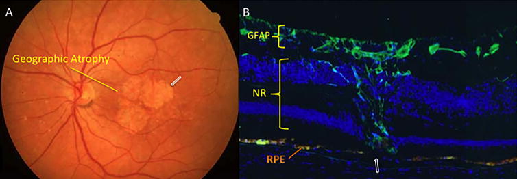
Figure 1.3: Geographic Atrophy
A) Fundal view of a patient with geographic atrophy, note the pale areas of retinal atrophy (arrow). B) Section of retina showing RPE cell loss (arrowed) with overlying loss of photoreceptors (PRL) at the macula. This area is replaced by intra-retinal glial tissue (anti-GFAP: green) (Courtesy of Dr Ann Milam, Scheie Eye Institute, University of Pennsylvania, Philadelphia, USA)
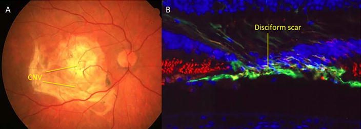
Figure 1.4: Advanced Neovascular Age-related Macular Degeneration
A) Fundal view of patient with advanced age related macular degeneration. A disciform scar is located at the macula. B) A immunofluorescence image of a macula with AMD, rods (red) surround an area of RPE and photoreceptor cell loss, replaced by a sub-retinal and intra-retinal glial (anti-GFAP: green) scar. (Courtesy of Dr Ann Milam, Scheie Eye Institute, University of Pennsylvania, Philadelphia, USA)


