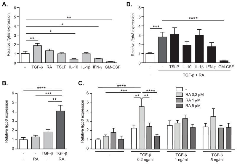
Figure 5. TGF-β and RA directly induce Itgb8 expression in spleen DCs.
CD11c+ spleen DCs were sorted with magnetic beads and then cultured in serum free medium for 16h. Cells were either left untreated (−) or stimulated with TGF-β, retinoic acid (RA), or both (A–C). In addition cells were treated with TSLP, IL-10, IL-1β, IFN-γ or GM-CSF, without (B) or with (D) addition of TGF-β and RA. 0.2 ng/ml TGF-β and 0.2 μM RA were used unless indicated otherwise. β8 integrin (Itgb8) gene expression was assessed by quantitative RT-PCR analysis relative to Actb and presented relative to levels in unstimulated spleen DCs. Histograms show mean ± SEM of cultures from 15 (A), 6 (B; D) or 9 (C) independent pools of mice from at least 3 separate experiments. *, p<0.05; **, p<0.005; ***, p<0.0005; ****, p<0.0001; one-way ANOVA with Dunnet’s post-hoc test (A–B; D) or two-way ANOVA with Tukey’s post-hoc test (C).

