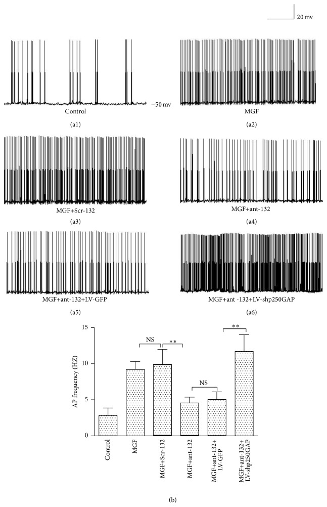
Figure 4.
Representative trace of AP in cultured hippocampal neurons. Cultured hippocampal neurons were separately transfected at DIV7 with Scr-132, ant-132, ant-132+LV-GFP, and ant-132+LV-shp250GAP and then treated with MGF medium at DIV10. AP frequency was tested 24 h after the MGF treatment. (a1) Representative recordings of non-MGF medium-treated neurons (control). (a2) Representative recordings of MGF medium-treated neurons. (a3–a6) Representative recordings of MGF medium-treated neurons separately after Scr-132, ant-132, ant-132+LV-GFP, and ant-132+LV-shp250GAP transfection. (b) Statistical analysis of AP frequencies. NS p > 0.05, ∗ p < 0.05, and ∗∗ p < 0.01 (n = 24; the data are representative of 5 experiments).

