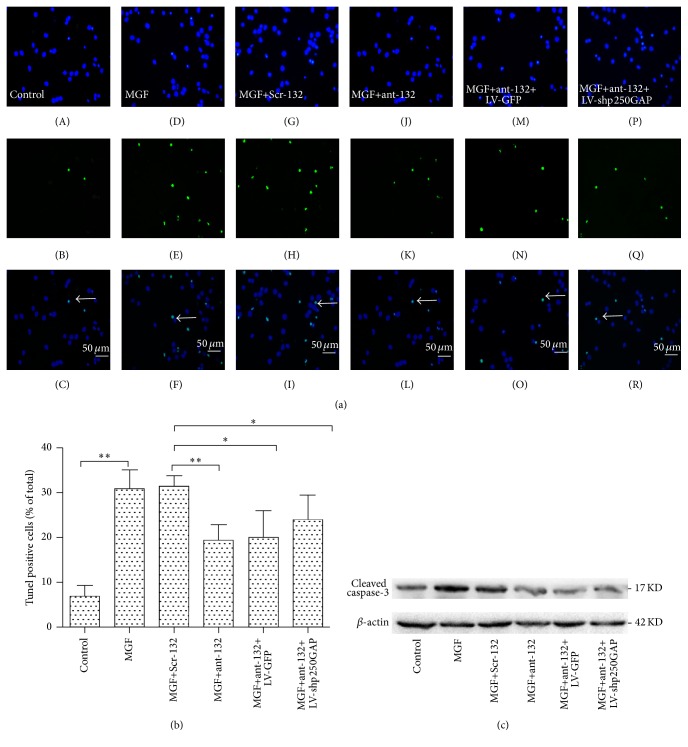
Figure 5.
(a) Neuroprotective effect of ant-132 in cultured MGF-treated hippocampal neurons. TUNEL assay was used to evaluate the apoptosis level. Apoptotic cells in the non-MGF-treated control group (A–C). MGF medium-treated control group (D–F). Neurons pretreated with Scr-132 (G–I). Neurons pretreated with ant-132 (J–L). Neurons pretreated with ant-132+LV-GFP (M–O). Neurons pretreated with ant-132+LV-shp250GAP (P–R). The white arrow indicates the apoptotic cell. Scale bar is 50 µm. (b) Histogram showing the percentage of cells with condensed nuclei in each group. The data are presented the mean ± SD values and subjected to ANOVA and Tukey's posttest. ∗ p < 0.05; ∗∗ p < 0.01 (n = 6; the data are representative of 5 experiments).

